3D Printing and Biofabrication by Aleksandr Ovsianikov James Yoo & Vladimir Mironov
Author:Aleksandr Ovsianikov, James Yoo & Vladimir Mironov
Language: eng
Format: epub
Publisher: Springer International Publishing, Cham
3 Inkjet-Mediated Gene Transfection and Inkjet Printing Biology Molecule
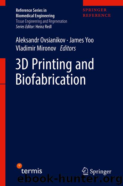
In addition to print cells, inkjet printing also have capacity to print proteins, cell guidance, and combination biologics. Several reports have demonstrated the interesting side effect of thermal inkjet printing technology and apply it to gene transfection and intracellular delivery (Xu et al. 2009b; Cui et al. 2010; Shattil et al. 1992; Owczarczak et al. 2012). Xu et al. firstly introduced a novel inkjet-mediated technology that gene transfection and cell delivery can be simultaneously achieved (Xu et al. 2009b). In this study, porcine aortic endothelial (PAE) cells and green fluorescent protein-coding (GFP) plasmids were co-printed into fibrin gel substrate. The co-printed plasmids could be transfected into cells and then expressed. They found that the viability of printed cells was over 90% and transfection efficiency was over 10%. The transfected cells could then be precise printed into predefined positions, and GFP could be expressed in in vitro and in vivo experiments. The author also postulated the mechanism of inkjet-mediated gene transfection. When cells and plasmids pass through the channel of printing head during the co-printing process, the high shear stress and heat may produce transient membrane pores. Plasmids can then get into the pores and expressed in cells (Fig. 4). Combining gene modification and cell delivery can benefit the field of tissue engineering and regenerative medicine, because it is important to facilitate the cell with certain function to form functional tissue and organ. Cui et al. further studied the thermal inkjet printing induced gene transfection (Cui et al. 2010). This study had a more comprehensive understanding on the influence of printing procedure on printed cells, such as cell viability and the size of cell membrane pores. Cell concentration was also optimized. Chinese hamster ovary (CHO) cells and fibrillarin- or GFP-fused plasmids were co-printed to achieve transfected cells, and it was observed that the transfection efficiency was above 30%, while cell viability was 89%. Furthermore, the study evaluated the membrane pore size and membrane repair time by incubating and staining the printed cells with dextran molecules (Shattil et al. 1992). Dextran can only penetrate the membrane pores, so the average Stokes diameters of the dextran molecules were used to indicate the size of membrane pores. Finally, it was observed that transient membrane pores can be repaired within 2 h. The study introduced that inkjet cell printing technology creates transient membrane pores during the printing process which holds possibility to be applied in intracellular delivery, such as genes, proteins, and factors’ transfection. Owczarczak et al. reported how to use a standard inkjet printer to process cells with fluorescent G-actin transfected (Owczarczak et al. 2012). Other researchers can follow their video instruction to convert a standard HP DeskJet 500 printer to an inkjet bioprinter. The printer has the capability to print cells and defined cellular microenvironments, leading to defined functional tissue structures.
Fig. 4(a) Schematic drawing of the postulated mechanism for inkjet-induced gene transfection. (b) The co-printed PAE cells after 2 days. (c) The green fluorescence expressed in co-printed cells.
Download
This site does not store any files on its server. We only index and link to content provided by other sites. Please contact the content providers to delete copyright contents if any and email us, we'll remove relevant links or contents immediately.
The Mikado Method by Ola Ellnestam Daniel Brolund(26278)
Hello! Python by Anthony Briggs(25205)
Secrets of the JavaScript Ninja by John Resig Bear Bibeault(24435)
Kotlin in Action by Dmitry Jemerov(23526)
The Well-Grounded Java Developer by Benjamin J. Evans Martijn Verburg(22869)
Dependency Injection in .NET by Mark Seemann(22658)
OCA Java SE 8 Programmer I Certification Guide by Mala Gupta(21420)
Algorithms of the Intelligent Web by Haralambos Marmanis;Dmitry Babenko(20258)
Grails in Action by Glen Smith Peter Ledbrook(19332)
Adobe Camera Raw For Digital Photographers Only by Rob Sheppard(17047)
Sass and Compass in Action by Wynn Netherland Nathan Weizenbaum Chris Eppstein Brandon Mathis(16357)
Secrets of the JavaScript Ninja by John Resig & Bear Bibeault(14071)
Test-Driven iOS Development with Swift 4 by Dominik Hauser(12245)
Jquery UI in Action : Master the concepts Of Jquery UI: A Step By Step Approach by ANMOL GOYAL(11520)
A Developer's Guide to Building Resilient Cloud Applications with Azure by Hamida Rebai Trabelsi(10637)
Hit Refresh by Satya Nadella(9212)
The Kubernetes Operator Framework Book by Michael Dame(8574)
Exploring Deepfakes by Bryan Lyon and Matt Tora(8424)
Robo-Advisor with Python by Aki Ranin(8366)
